Medical visualizations
Schematic medical visualizations of the heart and kidneys for a scientific journal illustration
The Problem
3D visual illustrations considerably enrich textual medical content: scientific presentations and medical articles. The value of illustration is also highly regarded by the authors who have the need for publishing their writings in journals, magazines, and websites. It is a proven fact: good illustrations immediately capture attention and settle in the viewers’ minds.
The Solution - Detailed Medical Visualizations
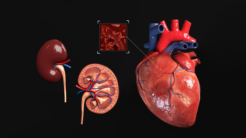
We worked on a schematic visual illustration of the heart and kidneys for various scientific medical magazines. This medical visualization depicts the detailed external and internal anatomy of the heart and kidneys. The anatomical heart structure includes the right and left anterior descending coronary arteries, auricles, ventricles, and great vessels (superior and vena cavae, pulmonary trunk, and aorta). The visualization also depicts a coronal section of the kidneys, so one can see their anatomy, which contains the major calyx, renal pelvis, ureter, renal cortex, renal columns, renal papilla, minor calyx, and renal pyramid. Done and delivered – 3D modeling, texturing, Blender Cycles materials, animation of the heart activity simulation, lighting, composition, and rendering.
Conclusion
Working alongside medical professionals is extremely gratifying. Together with doctors, surgeons, and other medical staff, we create impeccable, detailed-oriented images that not only inform but help to learn too.